TRANSLATIONAL ONCOLOGY GROUP
Research

Translational Oncology Group

Research

Research

Research programmes

Our research integrates chromatin biology, transcriptional regulation and functional models to address therapeutically relevant problems in ovarian and women's cancers.
01

H2Bub1 and chromatin remodelling in response to therapeutic DNA damage

How does H2B monoubiquitination (H2Bub1) redistribute after chemotherapy-induced DNA damage, and how do those changes control expression of genes that determine cell fate?

H2Bub1 is a co‑transcriptional chromatin mark that is globally reduced following platinum‑based DNA damage but becomes locally enriched at selected genes. Our work interrogates where and when H2Bub1 is deposited or removed after therapy, and how those local changes contribute to the activation or repression of genes involved in DNA repair, cell cycle arrest and cell death.

We combine defined DNA‑damage models and drug treatments with genome‑wide profiling to map H2Bub1 occupancy, correlate these changes with transcriptional outputs and test consequences for cell survival and sensitivity to chemotherapy in ovarian cancer models.
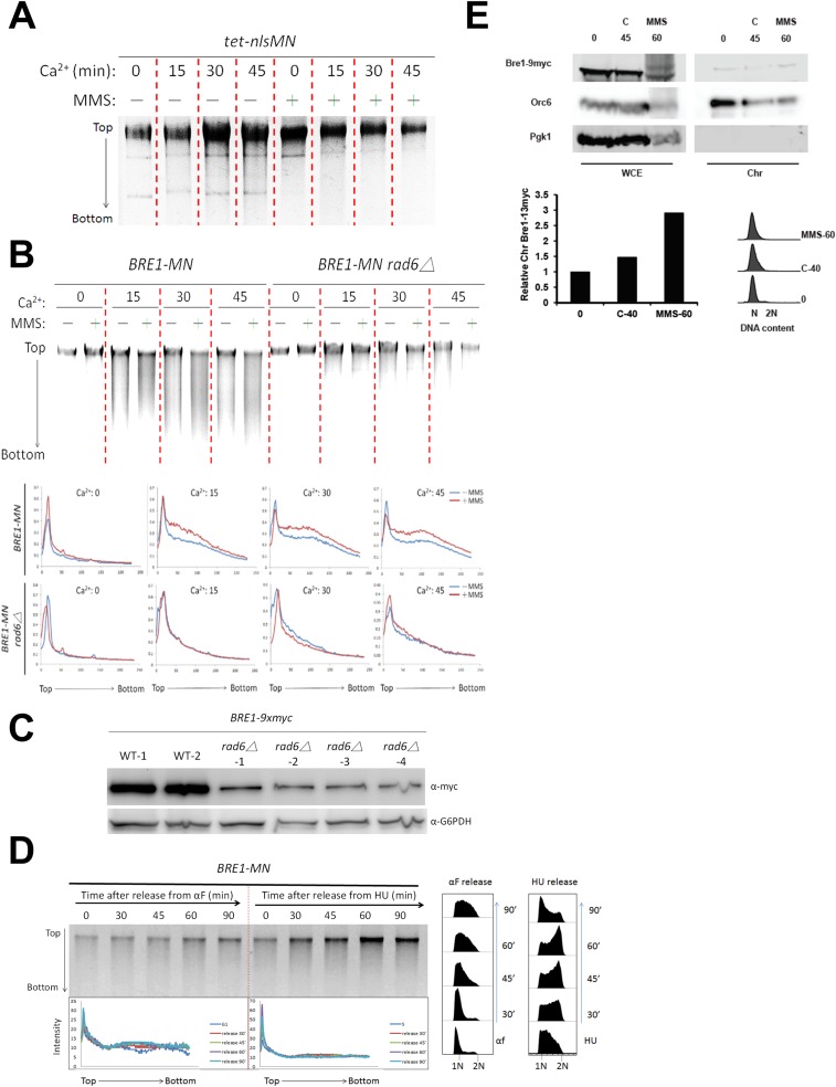
Figure(s) from: "Monoubiquitylation of histone H2B contributes to the bypass of DNA damage during and after DNA replication" — shows H2Bub1 dynamics and chromatin context relevant to DNA‑damage responses and redistribution of H2Bub1 after genotoxic stress.
02

p53 target‑gene regulation and chromatin control

How does p53 influence H2Bub1 and other chromatin marks to regulate target gene expression after genotoxic stress?

p53 is not simply a sequence‑specific transcription factor: it also interfaces with chromatin modifiers and the transcriptional machinery. Our group investigates how p53 status (wild‑type versus mutant) alters H2Bub1 deposition and downstream gene programmes that determine therapy outcomes.

We use isogenic cell lines and patient‑derived samples to link p53 activation states with chromatin mark redistribution, functional readouts of target gene activation (for example CDKN1A/p21, MDM2, BBC3/PUMA) and drug sensitivity, including routes to ferroptosis described in the literature where p53 can regulate H2Bub1 at key loci.

ChIP-seq / heatmap and UCSC genome-browser panels from the UTS-associated study showing H2Bub1 enrichment at p53 target loci after cisplatin (see Figure 1 and accompanying heatmaps in the article).
03

H2Bub1 loss, chromatin accessibility and ovarian cancer progression

What role does early loss of H2Bub1 play in ovarian tumour evolution, immune microenvironment changes and clinical outcome?

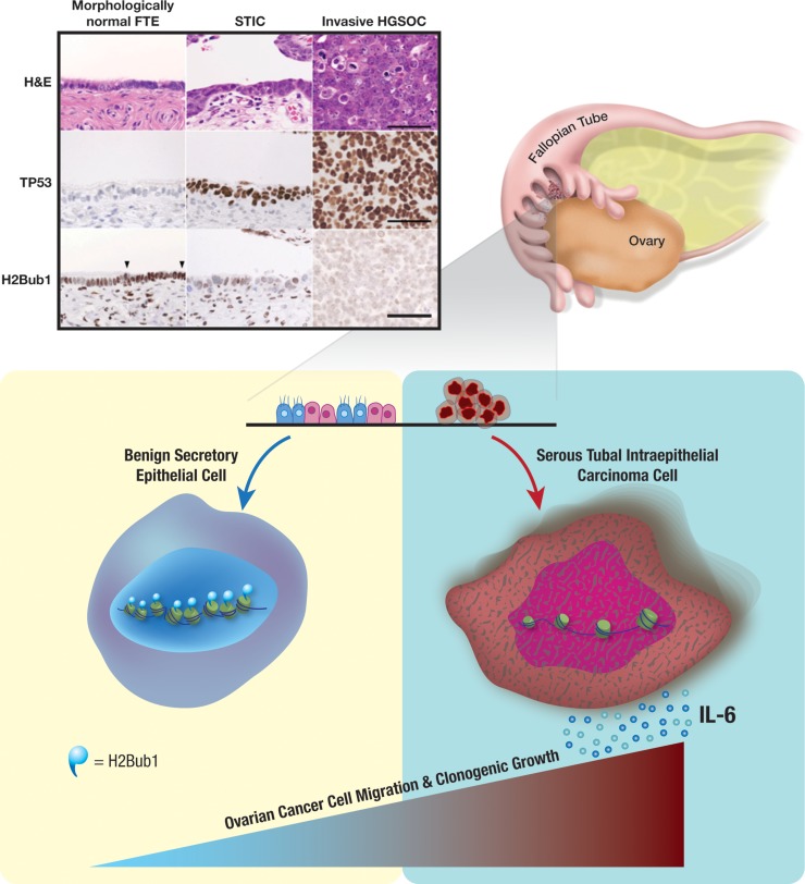
Multiple studies including work from our group show that H2Bub1 is frequently reduced or lost in ovarian tumours compared with normal fallopian tube epithelium. We test whether that loss promotes chromatin accessibility changes that increase pro‑tumour inflammation, alter immune infiltration and enable progression.

We integrate tissue‑based analyses (tissue microarrays, immunohistochemistry), ATAC‑seq for chromatin accessibility and in vitro modelling to define how H2Bub1 depletion changes gene regulatory landscapes and the tumour microenvironment in ovarian cancer.

Figures demonstrating p53-dependent changes in H2B monoubiquitination at the SLC7A11 regulatory region and functional links to ferroptosis (see Figure 4 and related panels in the article).
04

Mechanisms of drug resistance: links between DDR, chromatin and chemotherapy response

Which chromatin‑mediated mechanisms enable ovarian cancer cells to resist platinum‑based chemotherapy, and can these be targeted to restore sensitivity?

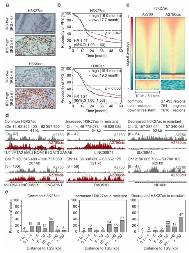
Resistance to platinum and related therapies remains the primary clinical barrier in high‑grade serous and other ovarian cancers. Our work explores how chromatin remodelling (including H2Bub1 loss), interaction with DDR factors and p53 status converge to permit tolerance or repair pathways that underlie resistance.

We seek to identify combinatorial vulnerabilities arising from altered chromatin states — for example altered recruitment of repair factors, shift to error‑prone repair, or immune signalling changes — and test targeted interventions that re‑sensitise resistant models to chemotherapy.

Figure(s) from: "Functional Analysis of Non-Genetic Resistance to Platinum in Epithelial Ovarian Cancer Reveals a Role for the MBD3-NuRD Complex in Resistance Development" — figures describing chromatin-mediated mechanisms associated with platinum resistance and candidate chromatin regulators.